924
Lectures Watched
Since January 1, 2014
Since January 1, 2014
- A History of the World since 1300 (68)
- History of Rock, 1970-Present (50)
- A Brief History of Humankind (48)
- Chinese Thought: Ancient Wisdom Meets Modern Science (35)
- The Modern World: Global History since 1760 (35)
- The Bible's Prehistory, Purpose, and Political Future (28)
- Introduction aux éthiques philosophiques (27)
- Jesus in Scripture and Tradition (25)
- Roman Architecture (25)
- Sexing the Canvas: Art and Gender (23)
- Descubriendo la pintura europea de 1400 a 1800 (22)
- Introduction aux droits de l'homme (19)
- Buddhism and Modern Psychology (18)
- Calvin: Histoire et réception d'une Réforme (17)
- The Ancient Greeks (16)
- À la découverte du théâtre classique français (15)
- The French Revolution (15)
- Letters of the Apostle Paul (14)
- Key Constitutional Concepts and Supreme Court Cases (14)
- Christianisme et philosophie dans l'Antiquité (14)
- Egiptología (12)
- Western Music History through Performance (10)
- The Rise of Superheroes and Their Impact On Pop Culture (9)
- The Great War and Modern Philosophy (9)
- Alexander the Great (9)
- Greek and Roman Mythology (9)
- Human Evolution: Past and Future (9)
- Phenomenology and the Conscious Mind (9)
- Masterpieces of World Literature (8)
- Villes africaines: la planification urbaine (8)
- Greeks at War: Homer at Troy (7)
- Pensamiento Científico (7)
- MongoDB for Node.js Developers (7)
- Fundamentos de la escritura en español (7)
- Introduction to Psychology (7)
- Programming Mobile Applications for Android (7)
- The Rooseveltian Century (6)
- Karl der Große - Pater Europae (6)
- Fake News, Facts, and Alternative Facts (6)
- Reason and Persuasion Through Plato's Dialogues (6)
- The Emergence of the Modern Middle East (6)
- A Beginner's Guide to Irrational Behavior (6)
- Lingua e cultura italiana: avanzata (6)
- L'avenir de la décision : connaître et agir en complexité (5)
- Understanding Einstein: The Special Theory of Relativity (5)
- Dinosaur Paleobiology (5)
- Exploring Beethoven's Piano Sonatas (5)
- War for the Greater Middle East (4)
- Emergence of Life (4)
- Introduction to Public Speaking (4)
- The Kennedy Half Century (4)
- Problèmes métaphysiques à l'épreuve de la politique, 1943-1968 (4)
- Designing Cities (4)
- Western Civilization: Ancient and Medieval Europe (3)
- Paleontology: Early Vertebrate Evolution (3)
- Orientierung Geschichte (3)
- Moons of Our Solar System (3)
- Introduction à la philosophie de Friedrich Nietzsche (3)
- Devenir entrepreneur du changement (3)
- La Commedia di Dante (3)
- History of Rock and Roll, Part One (3)
- Formation of the Universe, Solar System, Earth and Life (3)
- Initiation à la programmation en Java (3)
- La visione del mondo della Relatività e della Meccanica Quantistica (3)
- The Music of the Beatles (3)
- Analyzing the Universe (3)
- Découvrir l'anthropologie (3)
- Postwar Abstract Painting (3)
- The Science of Religion (2)
- La Philanthropie : Comprendre et Agir (2)
- Highlights of Modern Astronomy (2)
- Materials Science: 10 Things Every Engineer Should Know (2)
- The Changing Landscape of Ancient Rome (2)
- Lingua e letteratura in italiano (2)
- Gestion des aires protégées en Afrique (2)
- Géopolitique de l'Europe (2)
- Introduction à la programmation en C++ (2)
- Découvrir la science politique (2)
- Our Earth: Its Climate, History, and Processes (2)
- The European Discovery of China (2)
- Understanding Russians: Contexts of Intercultural Communication (2)
- Philosophy and the Sciences (2)
- Søren Kierkegaard: Subjectivity, Irony and the Crisis of Modernity (2)
- The Fall and Rise of Jerusalem (2)
- The Science of Gastronomy (2)
- Galaxies and Cosmology (2)
- Introduction to Classical Music (2)
- Art History for Artists, Animators and Gamers (2)
- L'art des structures 1 : Câbles et arcs (2)
- Russian History: from Lenin to Putin (2)
- The World of Wine (1)
- Wine Tasting: Sensory Techniques for Wine Analysis (1)
- William Wordsworth: Poetry, People and Place (1)
- The Talmud: A Methodological Introduction (1)
- Switzerland in Europe (1)
- The World of the String Quartet (1)
- Igor Stravinsky’s The Rite of Spring (1)
- El Mediterráneo del Renacimiento a la Ilustración (1)
- Science of Exercise (1)
- Социокультурные аспекты социальной робототехники (1)
- Russian History: from Lenin to Putin (1)
- The Rise of China (1)
- The Renaissance and Baroque City (1)
- Visualizing Postwar Tokyo (1)
- In the Night Sky: Orion (1)
- Oriental Beliefs: Between Reason and Traditions (1)
- The Biology of Music (1)
- Mountains 101 (1)
- Moral Foundations of Politics (1)
- Mobilité et urbanisme (1)
- Introduction to Mathematical Thinking (1)
- Making Sense of News (1)
- Magic in the Middle Ages (1)
- Introduction to Italian Opera (1)
- Intellectual Humility (1)
- The Computing Technology Inside Your Smartphone (1)
- Human Origins (1)
- Miracles of Human Language (1)
- From Goddard to Apollo: The History of Rockets (1)
- Hans Christian Andersen’s Fairy Tales (1)
- Handel’s Messiah and Baroque Oratorio (1)
- Theater and Globalization (1)
- Gestion et Politique de l'eau (1)
- Une introduction à la géographicité (1)
- Frontières en tous genres (1)
- Créer et développer une startup technologique (1)
- Découvrir le marketing (1)
- Escribir para Convencer (1)
- Anthropology of Current World Issues (1)
- Poetry in America: Whitman (1)
- Introducción a la genética y la evolución (1)
- Shakespeare: On the Page and in Performance (1)
- The Civil War and Reconstruction (1)
- Dinosaur Ecosystems (1)
- Développement durable (1)
- Vital Signs: Understanding What the Body Is Telling Us (1)
- Imagining Other Earths (1)
- Learning How to Learn (1)
- Miracles of Human Language: An Introduction to Linguistics (1)
- Web Intelligence and Big Data (1)
- Andy Warhol (1)
- Understanding the Brain: The Neurobiology of Everyday Life (1)
- Practicing Tolerance in a Religious Society (1)
- Subsistence Marketplaces (1)
- Physique générale - mécanique (1)
- Exercise Physiology: Understanding the Athlete Within (1)
- Introduction to Mathematical Philosophy (1)
- What Managers Can Learn from Great Philosophers (1)
- A la recherche du Grand Paris (1)
- The New Nordic Diet (1)
- A New History for a New China, 1700-2000 (1)
- The Magna Carta and its Legacy (1)
- The Age of Jefferson (1)
- History and Future of Higher Education (1)
- Éléments de Géomatique (1)
- 21st Century American Foreign Policy (1)
- The Law of the European Union (1)
- Design: Creation of Artifacts in Society (1)
- Introduction to Data Science (1)
- Configuring the World (1)
- From the Big Bang to Dark Energy (1)
- Animal Behaviour (1)
- Programming Mobile Services for Android Handheld Systems (1)
- The American South: Its Stories, Music, and Art (1)
- Care of Elders with Alzheimer's Disease (1)
- Contagious: How Things Catch On (1)
- Constitutional Law - The Structure of Government (1)
- Narratives of Nonviolence in the American Civil Rights Movement (1)
- Christianity: From Persecuted Faith to Global Religion (200-1650) (1)
- Age of Cathedrals (1)
- Controversies of British Imperialism (1)
- Big History: From the Big Bang until Today (1)
- Bemerkenswerte Menschen (1)
- The Art of Poetry (1)
- Superpowers of the Ancient World: the Near East (1)
- America Through Foreign Eyes (1)
- Advertising and Society (1)
Hundreds of free, self-paced university courses available:
my recommendations here
my recommendations here
Peruse my collection of 275
influential people of the past.
influential people of the past.
View My Class Notes via:
Receive My Class Notes via E-Mail:
Contact Me via E-Mail:
edward [at] tanguay.info
Notes on video lecture:
Basic Heart Anatomy
Notes taken by Edward Tanguay on May 22, 2014 (go to class or lectures)

Choose from these words to fill the blanks below:
sinus, atria, pulmonary, lungs, pericardium, thoracic, mediastinum, deoxygenated, thinner, lungs, thymus, membranes, thicker, pulmonary, upper, sternum
•
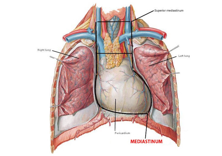
heart located right below
•
between two lungs
•
in cavity
•
enclosed by a membrane layer called the
•
lungs are also surrounded by membrane layers
•
it's very common in the body to have organs surrounded by which help to anchor the organs in position and protect them
•
located in the
•
contains the heart, the great vessels of the heart, the esophagus, the trachea, the phrenic nerve, the cardiac nerve, the thoracic duct, and the lymph nodes of the central chest
•
gland
•
glandular tissue over the heart
•
trachea with two branches
•
esophagus, passes behind the heart
•
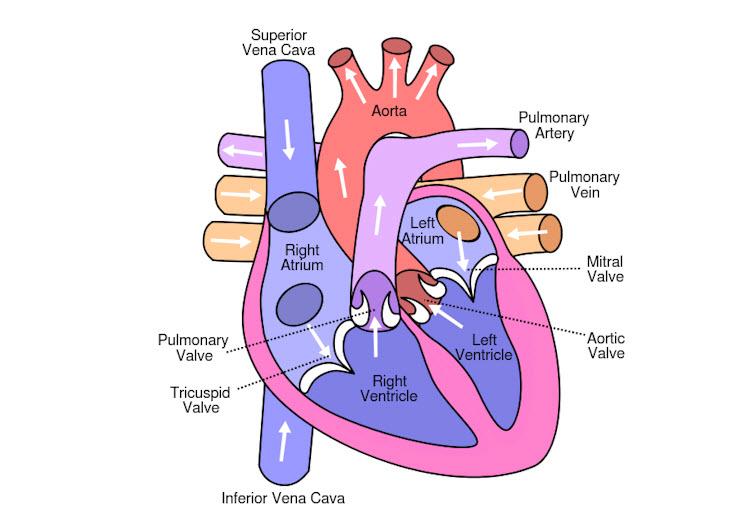
internal of heart
•
chambers
•
upper:
•
wall: interatria septrum
•
three vessels that carry blood back to the atrium
•
1. superior vena cava: from body
•
2. inferior vena cava: from lower body
•
3. coronary : blood from blood vessels from outside the wall of the heart itself
•
lower: ventricles
•
wall: interventricular septum
•
after blood has been in the right atrium, it passes into the right ventricle
•
contracts the blood into trunk
•
supplies the lungs with the blood to get oxygenated
•
blood comes back from the into the left atrium, then down to the left ventricle which pumps it out to the aorta
•
this is the beginning of the systemic circulation
•
systemic circulation: oxygenated blood to the body and back
•
circulation deoxygenated blood to the lungs and back
•
blood returns to the right atrium and the process is repeated
•
the walls of the atria are than the walls of the ventricles
•
because they have to push the blood out of the heart whereas the atria walls just have to push it to the ventricle below it
•
the walls of the left ventricle are because it pumps out to the whole body whereas the right only pumps to the
•
on an anatomy model, blue vessels carry deoxygenated, in reality this blood is less red, almost purplish
•
oxygenated blood as a bright red in color
Vocabulary:
| phrenic nerve, n. a nerve that originates in the neck and passes down between the lung and heart to reach the diaphragm, it is important for breathing ⇒ "The phrenic nerve is the sole motor supply to each hemidiaphragm." |
 mediastinum, n. [may-dee-AST-ri-num] region in mammals between the pleural sacs, containing the heart and all of the thoracic viscera except the lungs (vessels of the heart, esophagus, trachea, phrenic nerve, cardiac nerve, thoracic duct, lymph nodes of the central chest) ⇒ "Lesions in the posterior mediastinum may encroach on the esophagus, causing dysphagia or odynophagia." mediastinum, n. [may-dee-AST-ri-num] region in mammals between the pleural sacs, containing the heart and all of the thoracic viscera except the lungs (vessels of the heart, esophagus, trachea, phrenic nerve, cardiac nerve, thoracic duct, lymph nodes of the central chest) ⇒ "Lesions in the posterior mediastinum may encroach on the esophagus, causing dysphagia or odynophagia." |
| etiology, n. the study of causation, or origination ⇒ "With infectious etiologies, a widened mediastinum is a classic hallmark sign of anthrax infection." |
Spelling Corrections:
ventrical ⇒ ventricle
Flashcards:
membrane that surrounds the heart
pericardium
vessel that carries blood back from the upper body back into the heart
superior vena cava
on an anatomy model, blue vessels carry what kind of blood?
deoxygenated
blood comes back from the lungs into the left ventricle which pumps it out to the what?
aorta
what are the two kinds of circulation?
systemic (oxygenated blood to the body and back), and pulmonary (deoxygenated blood to the lungs and back)
which ventricle has a thicker wall and why?
the left, because it pumps out to the whole body whereas the right only pumps to the lungs